Spinal Fusion
Stabilising the spine for enhanced function and pain relief
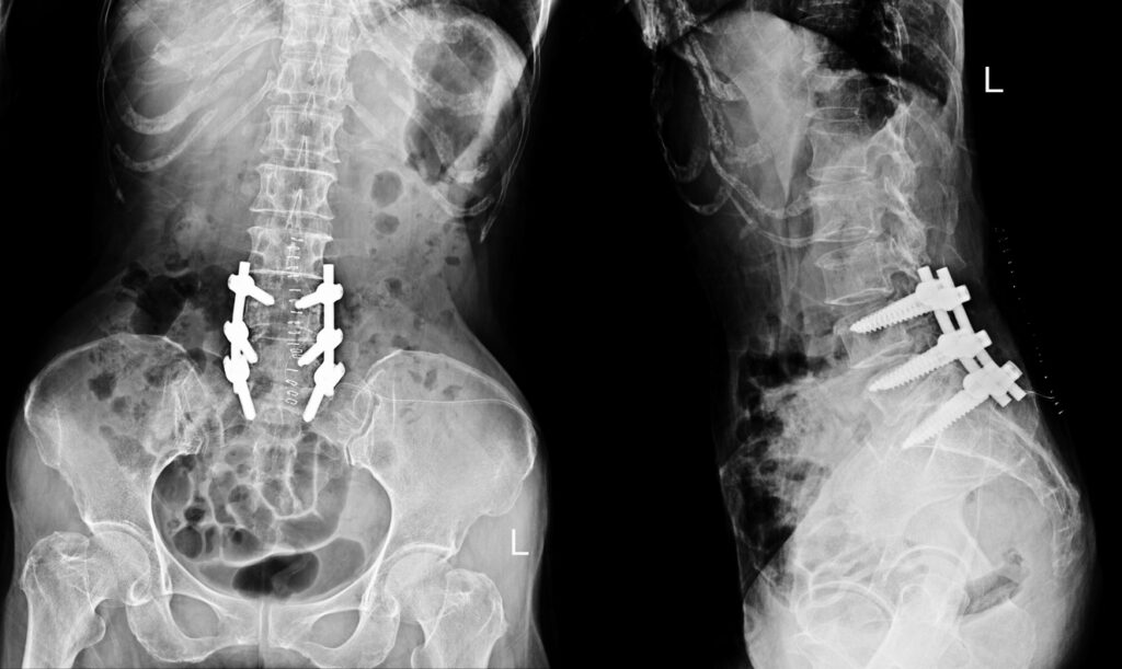
Spinal fusion is a surgical procedure designed to stabilise the spine by permanently joining two or more vertebrae. By eliminating movement between the affected vertebrae, the procedure can reduce pain, improve alignment, and restore stability to the spine. The surgery often involves the use of bone grafts, screws, or metal rods to encourage the vertebrae to heal together as a single, solid bone.
This approach may be recommended for a range of spinal conditions where instability, deformity, or persistent pain has not improved with non-surgical treatments. Common indications include degenerative disc disease, spinal instability, fractures, deformities such as scoliosis or kyphosis, and certain cases of spinal stenosis or herniated discs.
What is Spinal Fusion?
Spinal fusion is a procedure that joins two or more vertebrae together so they heal into a single, solid bone. By removing movement at the affected segment of the spine, the procedure helps reduce pain and improve spinal stability.
During surgery, the surgeon typically places a bone graft (natural or synthetic) between the vertebrae. Over time, this graft acts as a bridge, encouraging the vertebrae to fuse. Specialised implants such as screws, rods, or plates may also be used to hold the spine in position while the fusion takes place.
The exact technique can vary depending on the part of the spine being treated and the underlying condition. Spinal fusion can be performed in different regions of the spine; cervical (neck), thoracic (mid-back), or lumbar (lower back).
The main goal of spinal fusion is to relieve symptoms such as pain, weakness, or nerve irritation caused by instability, deformity, or degenerative changes in the spine.
Indications for Spinal Fusion Treatment
Spinal fusion may be recommended when conservative measures such as physiotherapy, medications, or injections have not adequately relieved symptoms. The procedure is generally considered for conditions that cause pain, instability, or deformity in the spine. Common indications include:
- Degenerative disc disease – when wear and tear of the spinal discs leads to persistent pain or instability.
- Spinal instability – often due to conditions such as spondylolisthesis, where one vertebra slips forward over another.
- Spinal stenosis – in cases where narrowing of the spinal canal causes nerve compression and symptoms are severe or persistent.
- Spinal deformities – including scoliosis or kyphosis, where fusion may help correct alignment and support stability.
- Trauma or fractures – to stabilise the spine following an injury.
- Spinal tumours or infections – in situations where removal of bone or tissue leaves the spine unstable and fusion is required for support.
The decision to undergo spinal fusion depends on the underlying diagnosis, the severity of symptoms, and the individual’s overall health. Your surgeon will carefully assess these factors and discuss whether spinal fusion is an appropriate option for your situation.
The Spinal Fusion Procedure
- Anaesthesia: The surgery is performed under general anaesthesia, ensuring patient comfort.
- Incision: A surgical incision is made, either on the back or the abdomen, depending on the location of the fusion.
- Bone Graft Placement: The surgeon prepares the vertebrae surfaces and places bone graft material between them to facilitate fusion.
- Instrumentation: Metal rods and screws may be used to secure the spine and enhance stability during the healing process.
- Closure: The incision is closed with sutures or staples, and a dressing is applied.
Recovery After Spinal Fusion
Recovery from spinal fusion varies depending on the underlying condition, the extent of the surgery, and individual health factors. The process is gradual, with improvement continuing for several months after surgery.
Immediately after surgery
- Patients usually spend several days in hospital to allow for monitoring and initial rehabilitation.
- Pain management, wound care, and support for early movement are provided by the hospital team.
- A physiotherapist will often guide safe techniques for getting in and out of bed, walking, and performing basic activities.
First few weeks at home
- Light walking is encouraged to promote circulation and healing.
- Activities that involve bending, twisting, or lifting are generally limited during this time.
- Patients may be fitted with a brace to support the spine as it heals.
- Follow-up appointments are scheduled to monitor progress and review imaging if needed.
Ongoing recovery
- Bone fusion typically takes several months, and during this time, activity levels are gradually increased.
- Physiotherapy may be recommended to strengthen surrounding muscles, improve mobility, and support long-term outcomes.
- Most people can return to desk-based work within 4–6 weeks, although more physically demanding roles may require a longer period of recovery.
Long-term Outcomes
- Full recovery can take 6–12 months, as the fusion continues to solidify.
- Many patients notice significant improvement in pain and stability, although flexibility in the fused segment of the spine will be reduced.
- Ongoing attention to posture, activity modification, and back care is important to protect other areas of the spine.
Risks and Complications
As with any surgery, spinal fusion carries certain risks. While complications are uncommon, it is important to be aware of them before deciding on treatment. Potential risks include:
- Infection at the surgical site
- Bleeding or blood clots
- Nerve injury leading to pain, numbness, or weakness
- Failure of the bones to fuse properly (non-union), which may require further surgery
- Hardware problems such as loosening or breakage of screws, rods, or plates
- Adjacent segment disease, where nearby spinal levels experience increased wear and tear due to altered biomechanics
- Persistent pain or stiffness, even after the surgery has healed
- Complications related to anaesthesia
Prof. Hunt will discuss these risks in detail and take careful steps to minimise them, including sterile surgical techniques, appropriate imaging, and close follow-up.