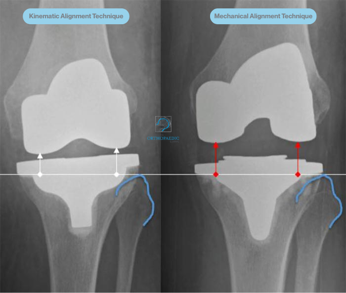
Kinematically aligned (KA), patient-specific total knee replacement is a modern, total knee replacement surgical technique designed to replicate the natural movement of your knee as closely as possible. Unlike traditional methods that follow a standardised mechanical alignment, the KA approach takes into account your individual bone structure, joint angles, and soft tissue balance. With KA, the goal is to position the implants in a way that mirrors how your knee functioned before arthritis developed, enhancing comfort, mobility, and overall satisfaction.
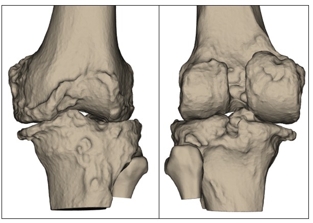
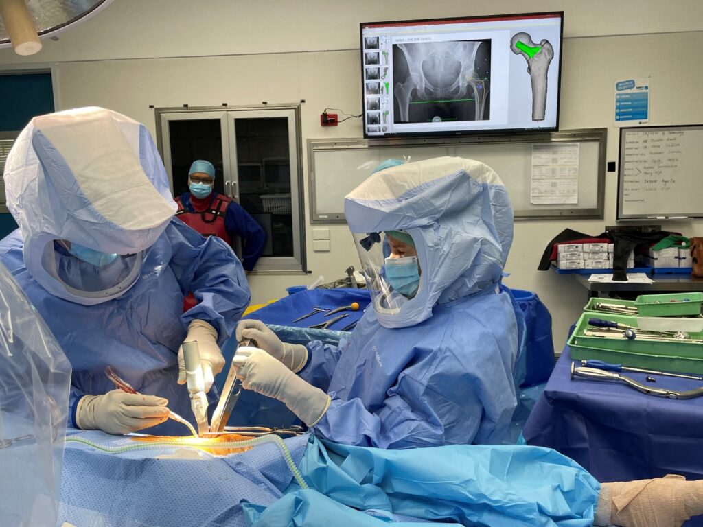
At Melbourne Orthopaedic Clinic, our orthopaedic surgeons use advanced preoperative imaging for total knee replacements, including CT scans to build a detailed 3D model of your knee. From this data, we create a surgical plan that is fully customised to your anatomy.
Where appropriate, patient-specific instrumentation (PSI) or computer-assisted navigation is also used to ensure each bone cut and implant position reflects your unique biomechanics. This level of precision may help reduce soft tissue disruption, support a more natural knee feel, and improve implant longevity.
Many patients report that their kinematically aligned total knee replacement feels more “normal” during everyday activities, particularly walking, climbing stairs, or kneeling. Because the joint is aligned to your individual anatomy rather than a generic axis, the surrounding ligaments are more likely to remain in balance, potentially resulting in less stiffness, better range of motion, and a smoother recovery.
A step-by-step guide to your surgery
Before your surgery, your orthopaedic team will use advanced imaging techniques such as X-rays and a CT scan to create a precise 3D model of your knee. This digital reconstruction allows your surgeon to assess the specific anatomy of your joint in detail, including bone alignment, the degree of wear from arthritis, and any irregularities in joint movement. This model forms the foundation for a fully personalised surgical plan. Every decision, from where bone resections are made to how the implants are aligned is guided by your own anatomy. By tailoring the procedure in this way, the aim is to optimise implant positioning, restore natural joint movement, and enhance the long-term function of your new knee.
To further enhance accuracy, your surgery may be performed using patient-specific instrumentation (PSI) or computer-assisted navigation, depending on your individual circumstances. These technologies use your preoperative scans to create customised surgical cutting guides or digital reference tools designed specifically for your knee. These guides are used during surgery to assist Prof. Hunt or Prof. Sallen in precisely removing bone and aligning the prosthetic components. This level of personalisation supports a more accurate fit and reduces the risk of malalignment, which may contribute to improved outcomes and implant longevity.
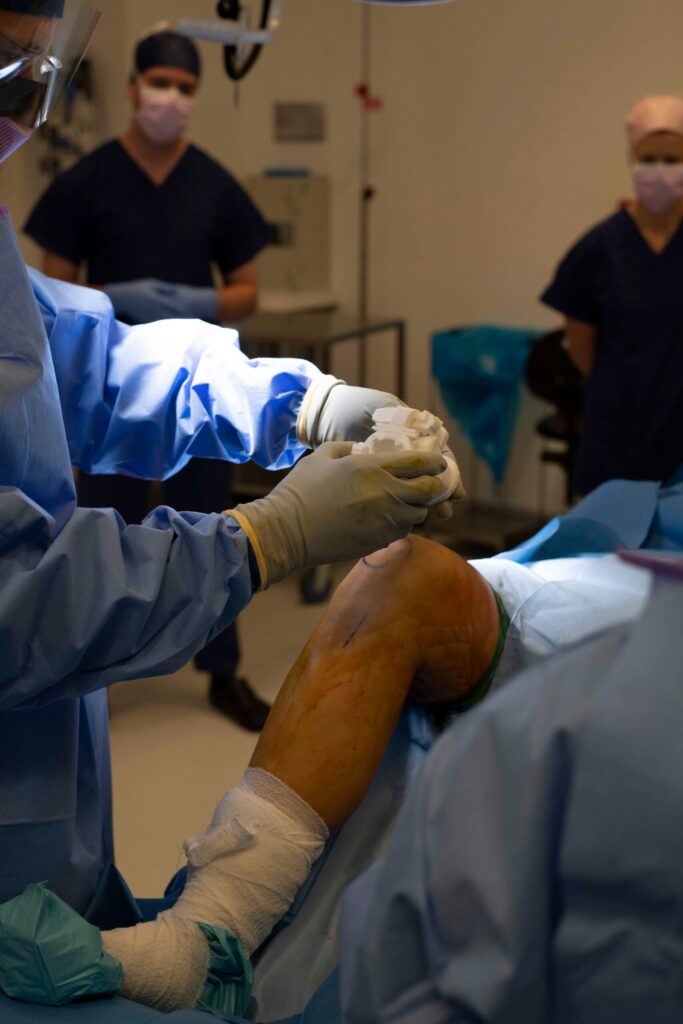
On the day of surgery, a carefully planned incision is made at the front of your knee to access the joint. The length of the incision will depend on your anatomy and the specific approach being used. Throughout this step, great care is taken to minimise trauma to the surrounding muscles, tendons, and soft tissues. Once the knee joint is visible, damaged cartilage and bone are carefully removed. Precision instruments and callipers are used to ensure that the exact amount of bone is taken away — just enough to accommodate the prosthetic components. The aim is to preserve healthy bone and tissue while preparing a stable surface for your implant.
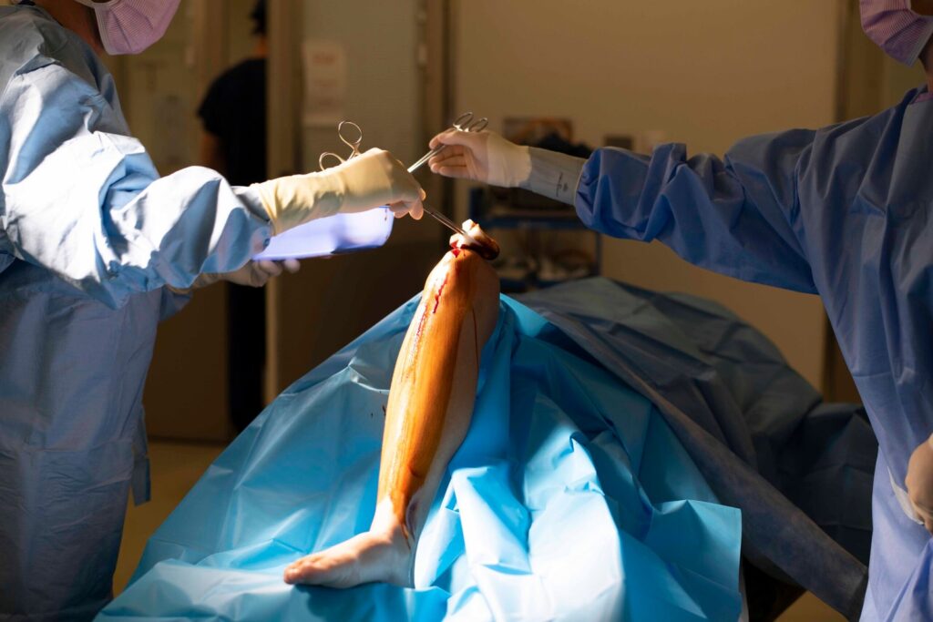
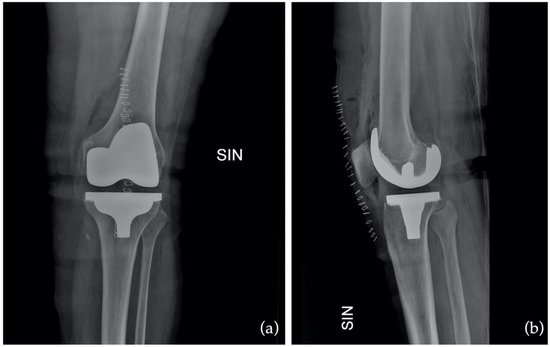
Your knee replacement involves inserting a set of prosthetic components — typically made from a combination of metal alloys and medical-grade plastic — to recreate the surfaces of the joint. These components are positioned to match the natural alignment and movement of your original knee, as closely as possible. In many cases, Prof. Hunt or Prof. Sallen may use computer-assisted techniques or patient-specific guides to help with positioning the implants with a high degree of precision. This helps restore natural joint kinematics and may contribute to improved function, better joint stability, and reduced wear over time.
Once the prosthetic components are in place and the joint is assessed for smooth movement and stability, the surgical site is thoroughly cleaned. The incision is then closed using sutures or staples, and a sterile dressing is applied to protect the wound. Care is taken to ensure the tissues are realigned properly, which supports optimal healing. You’ll be moved to the recovery area where your vital signs will be monitored as the anaesthetic wears off, and your postoperative care plan will begin shortly after.
A comprehensive listing of the orthopaedic services Prof. Justin Hunt & Prof. Vera Sallen provide including; joint replacements, sports injuries, trauma surgery, and minimally invasive procedures.
© Melbourne Orthopaedic Clinic 2026. All rights reserved.